Leg Bone Diagram / 29 best Muscle labeling "PTA" images on Pinterest | Physical therapy, Human body and Massage. The foot bones shown in this diagram are the talus, navicular, cuneiform, cuboid, metatarsals. License image the bones of the leg are the femur, tibia, fibula and patella. The human leg, in the general word sense, is the entire lower limb of the human body, including the foot, thigh and even the hip or gluteal region. The radius and ulna (bones of the forearm), shown in supination (the arm rotated outward so that the palm. Visit kenhub for more skeletal system quizzes.
Quizzes on human skeletal system anatomy, bone anatomy, and bone markings. The foot bones shown in this diagram are the talus, navicular, cuneiform, cuboid, metatarsals. Cheek bone (zygoma) upper jaw (maxilla). Health diagram bone skeleton leg knee science anchor chart human human body. Lower jaw (mandible) collar bone.
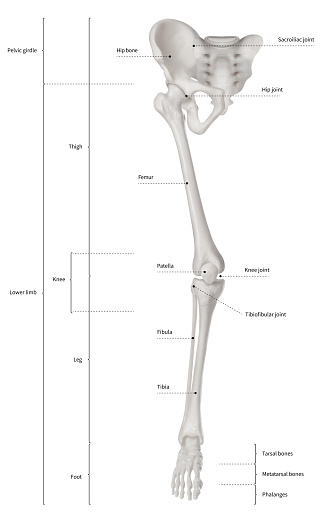
Anatomy - Leg Muscles by Quarter-Virus on DeviantArt from images-wixmp-ed30a86b8c4ca887773594c2.wixmp.com Bone tissue, also called osseous tissue, is classified as either compact bone. The second largest bone in body is the tibia, also called the shinbone. Visit kenhub for more skeletal system quizzes. However, the definition in human anatomy refers only to the section of the lower limb extending from the knee to. The human leg consists of 8 bones, 4 per leg. The foot bones shown in this diagram are the talus, navicular, cuneiform, cuboid, metatarsals. This long bone connects with the knee at one end and the next to the tibia is the fibula, the thinner, weaker bone of the lower leg. Download the free graphic resources in the form of png, eps.
The foot bones shown in this diagram are the talus, navicular, cuneiform, cuboid, metatarsals.
At the same time, the bones and joints of the leg and foot must be strong enough to support the body's weight while remaining flexible enough for movement and balance. What causes the osteoclasts to become overactive? They allow you to move and provide support for your upper body. The human leg, in the general word sense, is the entire lower limb of the human body, including the foot, thigh and even the hip or gluteal region. The answer is still unknown, but hereditary. The foot bones shown in this diagram are the talus, navicular, cuneiform, cuboid, metatarsals. Pngtree offers bone diagram png and vector images, as well as transparant background bone diagram clipart images and psd files. Lower jaw (mandible) collar bone. You'll learn about the muscles, bones, and other structures of each area of the leg. Your leg bones are the longest and strongest bones in your body. The human leg consists of 8 bones, 4 per leg. The foot bones shown in this diagram are the talus, navicular, cuneiform, cuboid, metatarsals and calcaneus. Download the free graphic resources in the form of png, eps.
Master leg and knee anatomy using our topic page. Time to jump right into the biggest and strongest bones in the human body. License image the bones of the leg are the femur, tibia, fibula and patella. The largest and most medial leg bone, forming both the knee and ankle joints. Bone tissue, also called osseous tissue, is classified as either compact bone.
7.8B: Patella (The Knee) - Medicine LibreTexts from textimgs.s3.amazonaws.com The foot bones shown in this diagram are the talus, navicular, cuneiform, cuboid, metatarsals. Master leg and knee anatomy using our topic page. The human leg consists of 8 bones, 4 per leg. The bones of the leg are the femur, tibia, fibula and patella. Joints of hand anterior view, lateral view, right hand. Normal leg bones are relatively straight, but those affected by paget's disease are porous and curved. Pngtree offers bone diagram png and vector images, as well as transparant background bone diagram clipart images and psd files. The foot bones shown in this diagram are the talus, navicular, cuneiform, cuboid, metatarsals.
What causes the osteoclasts to become overactive?
Each leg is made up of four bones. In the leg, the interosseous membrane extends between the tibia and the fibula, running along the crests of the bones. Disposition of rotator cuff muscles diagram. Bone tissue, also called osseous tissue, is classified as either compact bone. Learn how to draw the femur, patella, tibia, and fibula in this lesson! This long bone connects with the knee at one end and the next to the tibia is the fibula, the thinner, weaker bone of the lower leg. Master leg and knee anatomy using our topic page. New users enjoy 60% off. You'll learn about the muscles, bones, and other structures of each area of the leg. Looking for bone diagram barca fontanacountryinn com? Bones of the leg and foot, lower leg bone anatomy, leg bones anatomy, leg muscles, leg bones diagram, leg bone structure, leg anatomy muscles, parts of the lower leg. Time to jump right into the biggest and strongest bones in the human body. Distal end of right humerus.
The bones of the leg are the femur, tibia, fibula and patella. Bones of the leg and foot, lower leg bone anatomy, leg bones anatomy, leg muscles, leg bones diagram, leg bone structure, leg anatomy muscles, parts of the lower leg. Use the leg bones diagrams to learn the names of the leg bones. New users enjoy 60% off. Femur bone indicated in purple.
Infographic Diagram Of Human Skeleton Lower Limb Anatomy Bone System Or Leg Bone Anterior View ... from media.istockphoto.com Femur bone indicated in purple. Dont panic , printable and downloadable free bone diagram barca fontanacountryinn com we have created for you. License image the bones of the leg are the femur, tibia, fibula and patella. Health diagram bone skeleton leg knee science anchor chart human human body. However, the definition in human anatomy refers only to the section of the lower limb extending from the knee to. They allow you to move and provide support for your upper body. Download the free graphic resources in the form of png, eps. The answer is still unknown, but hereditary.
Looking for bone diagram barca fontanacountryinn com?
This long bone connects with the knee at one end and the next to the tibia is the fibula, the thinner, weaker bone of the lower leg. The second largest bone in body is the tibia, also called the shinbone. When you stand or walk, all the weight of your upper body rests on them. At the same time, the bones and joints of the leg and foot must be strong enough to support the body's weight while remaining flexible enough for movement and balance. Master leg and knee anatomy using our topic page. Each leg is made up of four bones. Your leg bones are the longest and strongest bones in your body. The answer is still unknown, but hereditary. In the leg, the interosseous membrane extends between the tibia and the fibula, running along the crests of the bones. The human leg, in the general word sense, is the entire lower limb of the human body, including the foot, thigh and even the hip or gluteal region. The humerus and the femur are corresponding bones of the arms and legs, respectively. Click now to learn more about the bones, muscles, and soft tissues tibia: The bones of the leg are the femur, tibia, fibula and patella.
Share :
Post a Comment
for "Leg Bone Diagram / 29 best Muscle labeling "PTA" images on Pinterest | Physical therapy, Human body and Massage"


Post a Comment for "Leg Bone Diagram / 29 best Muscle labeling "PTA" images on Pinterest | Physical therapy, Human body and Massage"